Oocyte I definition
Oocyte I designates a female germline diploid cell that forms during the cycle called oogenesis. Mammalian oocytes I enter phase I of meiosis during fetal development.
Oocyte I development
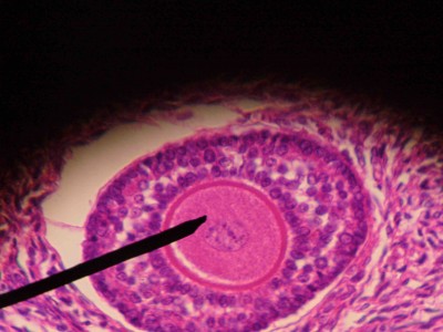
Oocyte I develops during oogenesis, a cycle through which oogonium develop and produce ovum. This cycle begins in fetal development, where primordial germ cells migrate to gonadal ridge and give rise to oogonium. Even during fetal development, oogonium progress through cell cycle to prophase I. The phase in which meiosis is interrupted is called dipotene and the oocyte is called oocyte I – diploid cell (figure 1). Oocyte I remains in prolonged prophase I from the fetal period to puberty.

Figure 1 – Histological image of a primary follicle. The needle points to oocyte I, inside the follicle.
In humans, the cycle remains postponed for decades (up to 12/13 years in women), and in the case of rats, this suspension lasts only months. Interruption of meiosis in prophase I and its postponement is only possible due to activity decrease of Cdk1/Cyclic B kinase complex (complex which exhibits high activity during a specific stage of meiosis and which is responsible for the passage from this stage to the next). Cyclic AMP is mainly responsible for inactivation of this complex, being produced by the oocyte itself.
When puberty is reached, there are peaks of luteinizing hormone (LH), which is responsible for releasing oocyte I from prophase I, causing it to progress in cycle. Progression in meiosis in response to LH succinctly involves cyclic GMP concentration reduction, which, consequently, leads to a cyclic AMP concentration decrease. Therefore, there is an increase in Cdk1/Cyclic B complex activity, which leads to release and progression in meiosis.
Oocyte I then proceeds by meiosis I, undergoing various modifications until first meiosis is complete. After this event, oocyte I (diploid cell) gives rise to two daughter cells (haploid cells): one becomes the oocyte II and the other forms the first polar globule.
References:
Abrieu A., Dorée M., Fisher D. (2001). The interplay between cyclin-B–Cdc2 kinase (MPF) and MAP kinase during maturation of oocytes. Journal of Cell Science 114, 257-267. The Company of Biologists Ltd.
Gilbert S. F. (2013). Developmental Biology (10ª edição). Sinauer Associates, Inc., Sunderland (MA).
Mandelbaum, J. (2000). Oocytes. Human Reproduction, 15, 11-18.
Von Stetina, J. R., & Orr-Weaver, T. L. (October de 2011). Developmental Control of Oocyte Maturation and Egg Activation in Metazoan Models. Cold Spring Harb Perspect Biology, 3(10), 19.


