Interphase definition
Interphase refers to the phase between two successive mitoses, during which chromosomes are relaxed and functionally active and where an intense metabolic activity takes place. It is the longest phase of cell cycle and is subdivided into G1 stage, S stage and G2 stage.
Cell Cycle
Cell cycle is divided into two successive phases: interphase, where duplication of genetic material and cellular constituents occurs, and phase M, where genetic material, cell constituents and cytoplasm are distributed.
Interphase characteristics
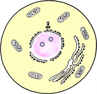
Cell stays 95% of cell cycle in the interphase, for example a human cell in proliferation has a 24h cell cycle, of which 23h are passed in interphase. At this stage, chromosomes are decondensed and randomly distributed through cell nucleus (figure 1), which has a morphologically uniform appearance when viewed under an optical microscope (figure 2). It is at this stage that DNA and cell constituents’ duplication and cell growth (doubling of size) occur; i.e., it is at this stage that cell performs all the processes necessary for cell cycle next phases.
G1
It concerns the interval (Gap – G) between mitosis and the onset of DNA replication. At this interval, cell is metabolically active and is continually growing, however has not yet begun to duplicate its DNA.
S
Synthesis stage, i.e., phase in which DNA replication occurs. This replication is semi-conservative, i.e., one of double-stranded DNA strand serves as a template for cell’s replication machinery to synthesize a new DNA strand. At the end, two chromosomes are obtained, each with two equal sister chromatids.
G2
Interval between synthesis phase and the beginning of M phase. In this interval, cell is synthesizing proteins needed to enter mitosis. DNA duplication is now complete, but cell continues to grow.

Figure 1 – Cell interphase schematic image.

Figure 2 – Interphase whitefish cell; Optical microscopy image.
Regulation
Cell progress from one phase to the next one in cell cycle is highly regulated to ensure that cells with excessive or missing DNA content are not perpetuated.
G1 checkpoint
An important cell cycle checkpoint occurs in G1, in which cell evaluates nutrients availability for the entire cell cycle duration, DNA integrity and proliferation signals that are or are not being sent by neighbour cells. If there are not enough nutrients or there are no signals from neighbour cells, cell enters in a resting phase where cell cycle does not progress – G0. This G0 stage is known to be the quiescent phase of cell cycle, in which cell can stay for long periods of time, without proliferating and not growing, with a very low metabolic rate.
This checkpoint functions as a restraint point: if all conditions for cycle progress are met, cell progresses to S stage, unable to stop until cycle ends. That is, from the moment cell progresses to S stage, it is committed to terminating cell cycle.
p53 protein is one of those responsible for stopping the cycle at this control point, which has its expression induced when DNA damage occurs.
G2 checkpoint
Another checkpoint occurs at G2, in which cell verifies if all DNA is correctly replicated; i.e., if all DNA was replicated and if it was only one time. If a signal is being sent by unreplicated DNA, cell stops cell cycle and does not advance to mitosis until all DNA is correctly duplicated. Once all DNA is replicated, cell progresses in cell cycle and progresses to mitosis.
At this check point, cell also evaluates whether DNA is damaged. If this occurs, cell delays the cycle long enough to be able to repair the DNA.
Study of cells in interphase
Cells in different stages of interphase are only distinguished by biochemical criteria. For example, cells in S stage are replicating DNA, so incorporate thymines at a fairly high rate. Thus, when radioactive thymines are added to proliferating cell culture medium, those in the S stage will incorporate these radioactive thymines, which will be visualized by autoradiography.
Cells in different interphase stages can also be distinguished by their content in DNA. For example, human cells in G1 are diploids (2n); cells in S stage, are replicating their DNA, i.e., are switching from diploids to tetraploids (2n to 4n); and G2 cells are tetraploids (4n). This DNA content is visualized after incubating the cells with a dye that binds to DNA and analyzed the cells by flow cytometry.
References:
Alberts B., Johnson A., Lewis J., Raff M., Keith R., Walter P. (2007). Molecular Biology of the Cell (5th edition). Garland Science, New York.
Cooper G.M. (2000). The Cell: A Molecular Approach (2th edition). Sinauer Associates, Sunderland (MA).
Griffiths A.J.F., Miller J.H., Lewontin R.C., Gelbart W.M. (1999). Modern Genetic Analysis (2nd edition). W. H. Freeman, New York.
Lodish H., Berk A., Zipursky S.L., Matsudaira P., Baltimore D., Darnell J. (2000). Molecular Cell Biology (4th edition). W. H. Freeman, New York.


