Chromatin Definition
Chromatin refers to a complex formed by DNA, histones and other proteins, which makes it possible for the eukaryotic cell to condense and store all its DNA inside the nucleus. Chromatin forms chromosomes.
Chromatin and Chromosomes
For its cycle (growth, metabolism, differentiation, division and death), eukaryotic cell needs a lot of information, all of which is stored in the form of desoxyribonucleic acid – DNA. Each eukaryotic cell has about 2m of DNA packed into a nucleus of 5-8um in diameter. For this to be possible it is necessary to condense and pack all the DNA into chromosomes. Human cells, for example, have 24 different pairs of chromosomes, i.e., 46 chromosomes. Each chromosome is composed of a long double-stranded DNA chain associated with histones and non-histone proteins that lead to the formation of a condensed configuration of the DNA molecule. This complex is called ‘chromatin’.
Chromatin’s characteristics
Chromatin usually contains twice as many proteins (histones and non-histones) compared to its amount of DNA, however, the histone mass is almost equal to the mass of DNA in the nucleus
Histones
Histones represent the largest class of proteins in chromatin and are small proteins that have a high content in positively charged amino acids – arginine and lysine. The presence of these amino acids makes it easier to bind the histones to the negatively charged DNA molecule. There are 5 different histones present in the chromatin: H1, H2A, H2B, H3 and H4.
Non-histone proteins
Non-histone proteins are regulatory proteins that are associated with specific DNA sequences. These proteins are, mainly, transcription factors, however, little is known about their function in the packing of chromatin.
Nucleosome
Histones associated with DNA form the first level of chromatin compaction, the nucleosome, which is characterized by 11nm fibers. At this level of compaction, the chromatin, viewed under the electron microscope, has the appearance of a pearl necklace (figure 1), the beads being the nucleosome and the necklace strand the DNA. Each nucleosome consists of a complex of eight histones – two molecules of H2A, two molecules of H2B, two molecules of H3 and two molecules of H4 – and by 146 nucleotides coiled around this complex. Each nucleosome is separated from the next by 80 nucleotides.

Figure 1 – DNA associated with proteins, forming the nucleosome, in the first level of chromatin compaction (11nm fibers). Optical microscopy image, where white arrows show DNA strands between nucleosomes and black arrows show nucleosomes.
The next level of chromatin compaction involves the condensation of this ‘beads on a string’ to form fibers of 30nm diameter. This higher level of compaction is achieved by the action of histone H1, which is bound to the nucleosome and ‘forces’ the DNA to bend and change its direction as it leaves the nucleosome, helping to form loops in the DNA and thus increasing its compaction.
Chromatin condensation levels
The condensation state of the chromatin and consequently of the chromosomes varies, depending on the phase of the cell cycle in which eukaryotic cell is found. During interphase, the cell cycle phase in which eukaryotic cell is doubling its genome and its constituents, most chromatin is in decondensed state of 30nm fibers – euchromatin – and 10% is in a highly condensed state – heterochromatin. In this phase of the cycle, chromosomes are called ‘interphase chromosomes’ (figure 2).
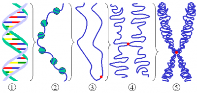
When eukaryotic cell enters in metaphase, the phase of the cell cycle where the cell prepares to divide the genomic material into two daughter cells, chromatin is all in the state of greatest compaction. At this stage, chromosomes are called ‘mitotic chromosomes’ and chromatin is highly condensed in 1400nm fibers (figure 2), and it is even possible to observe them through the optical microscope. This high compaction state makes replication and transcription impossible, but ensures that no piece of DNA molecule is lost during separation of sister chromatids to the different daughter cells.

Figure 2 – Different levels of chromatin compaction. 1) DNA molecule; 2) ‘beads on a string’, 11nm fibers; 3) 30nm fibers; 4) 700nm fibers and; 5) 1400nm fibers, mitotic chromosome.
Chromatin’s types in interphase
Euchromatin
During interphase it is necessary that chromatin is in its most decondensed state – euchromatin – to be possible to replicate and transcribe DNA. It should be noted that in DNA zone where genes are under active transcription, chromatin is not in the form of 30nm fiber, but rather in the conformation of ‘beads on a string’ (11nm fibers).
Heterochromatin
During interphase there are areas of chromatin that maintain their highly condensed conformation – heterochromatin. The conformation of heterochromatin does not allow the transcription of DNA located in these zones and, therefore, is located in zones of DNA that do not contains genes. This form of chromatin is located in areas of highly repetitive DNA, such and centromere and telomeres.
References:
Alberts B., Johnson A., Lewis J., Raff M., Keith R., Walter P. (2007). Molecular Biology of the Cell (5th edition). Garland Science, New York.
Cooper G.M. (2000). The Cell: A Molecular Approach (2th edition). Sinauer Associates, Sunderland (MA).
Lodish H., Berk A., Zipursky S.L., Matsudaira P., Baltimore D., Darnell J. (2000). Molecular Cell Biology (4th edition). W. H. Freeman, New York.


