Nucleosome definition
Nucleosome refers to the fundamental structural unit of chromatin in the eukaryotic cell. It is this protein complex that gives the chromatin the ‘necklace of beads’ aspect.
Cell nucleus and chromatine
The cell stores all information necessary for its growth, metabolism and division into the nucleus, in the form of DNA. However, if DNA molecule were in a relaxed and ‘stretched’ state, the nucleus would not be large enough to accommodate 2m of DNA in each cell. In order to solve this problem of space, cell made possible the packaging of DNA through formation of chromatin. Chromatin is characterized by existence of protein complexes (histones and other proteins) around which DNA coils, which under microscope is observed as a ‘bead on a string’. With this compaction the DNA is one third of its initial size.
Nucleosome characteristics
Nucleosome was discovered in 1974 after chromatin digestion with nucleases, which ‘cut’ DNA between nucleosomes.
Each nucleosome consists of a complex with about 10 nm, consisting of 8 histones (two histone molecules H2A, H2B, H3 and H4) and 146 nucleotides of DNA. Histones form a protein complex – octamer of histones – which lies at the center and around which 146 nucleotides of DNA are wound. Nucleosomes are separated by a small sequence of DNA that is at most up to 80 nucleotides.
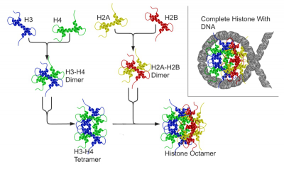
Histone octamer formation is done initially by formation of tetramers H2A-H2B and H3-H4. Subsequently, the two tetramers associate and form an octamer (figure 1). DNA binds to histone octamer through hydrogen bonds, hydrophobic interactions and electrostatic interactions. This linkage between DNA and histone octamer is facilitated/hampered by the presence of specific proteins.

Figure 1 – Schematic image of nucleosome three-dimensional structure. Histones are present in dimers; dimers H2A and H2B form a tetramer; dimers H3 and H4 form another tetramer; in turn, the two tetramer joins together and forms histone octamer. To the right of the image, histone octamer is observed centrally, with DNA wrapped around it.
References:
Alberts B., Johnson A., Lewis J., Raff M., Keith R., Walter P. (2007). Molecular Biology of the Cell (5th edition). Garland Science, New York.
Berg J.M., Tymoczko J.L., Stryer L. (2002). Biochemistry (5th edition). W. H. Freeman, New York.
Brown T.A. (2002). Genomes (2nd edition). Wiley-Liss, Oxford.
Cooper G.M. (2000). The Cell: A Molecular Approach (2th edition). Sinauer Associates, Sunderland (MA).
Griffiths A.J.F., Miller J.H., Lewontin R.C., Gelbart W.M. (1999). Modern Genetic Analysis (2nd edition). W. H. Freeman, New York.
Griffiths A.J.F., Miller J.H., Suzuki D.T., Lewontin R.C., Gelbart W.M. (2000). An Introduction to Genetic Analysis (7th edition). W. H. Freeman, New York.
Lodish H., Berk A., Zipursky S.L., Matsudaira P., Baltimore D., Darnell J. (2000). Molecular Cell Biology (4th edition). W. H. Freeman, New York.


