Concept of Prophase
The term prophase designates one of the subphases of mitosis, characterized by the reorganization of the cytoskeleton, migration of centriole to opposite cell poles, the appearance of the mitotic spindle, condensation of chromosomes, fragmentation of nuclear envelope and disappearance of the nucleolus.
Cell Cycle
The cell cycle is the basis of all life. The unicellular organisms use the cell cycle to reproduce and the multicellular to form the tissues and organs of the complex organism. This cycle is divided into Interphase and M phase. The first phase of the cycle, Interphase, is subdivided into phase G1, S phase and G2 phase, where duplication of genetic material and cellular components occurs. M-phase is subdivided into Mitosis – which includes Prophase, Prometaphase, Metaphase, Anaphase and Telophase – and Cytokinesis, where the nucleus and cytoplasm divide. At the end of a cell cycle, two cells exactly equal to its progenitor are formed.
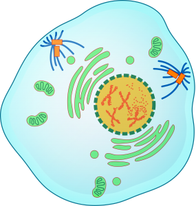
Features of Prophase (figure 1)
Mitotic Spindle
During Prophase, the reorganization of cytoskeleton occurs to initiate the formation of the mitotic spindle. The mitotic spindle is composed of microtubules and associated motor proteins, forming parallel to the axis of the cell in order to connect the opposing poles of the cell.
The formation of the spindle is abruptly triggered by cellular signals, which cause the long interphase microtubules to disappear and to form interpolar microtubules, kinetochore microtubules, and astral microtubules. These microtubules polymerize from the centrosome, a complex initially formed by a centriole that is duplicated during the interphase; being, in this phase, formed by two centrioles. The two centrioles separate and move to opposite locations of the nucleus. The polarity and dynamic instability of the microtubule polymerization from the centrioles that occurs at this stage of the cell cycle cause them to polymerize in opposite directions of the cell thus forming opposite poles of the mitotic spindle.
Chromosomes
At the same time that the mitotic spindle begins to form, the replicated chromosomes in the interphase, begin to condense within the nucleus. In this phase, the chromosomes’ chromatin is in the conformation of fibers of 30nm, which already makes possible the visualization of the chromosomes using the optical microscope (in a configuration like wire). The sister chromatids are linked via the centromere, which will serve as the binding site for the spindle microtubules in the next phase of the cell cycle.
Nucleus
The nuclear envelope consists of lamins that, with the onset of Prophase, are phosphorylated and lose affinity with each other, which leads to its disintegration. This fragmentation of the nuclear envelope makes it possible to attach the spindle microtubules to the kinetochore of the chromosomes, as explained above.
However, fragmentation of the nuclear envelope is not universal to all organisms. Yeast, for example, performs nuclear division during mitosis without fragmenting its nuclear envelope.

Figure 1 – Representative diagram of the various cellular changes that occur during Prophase.
References:
Alberts B., Johnson A., Lewis J., Raff M., Keith R., Walter P. (2007). Molecular Biology of the Cell (5th edition). Garland Science, New York.
Cooper G.M. (2000). The Cell: A Molecular Approach (2th edition). Sinauer Associates, Sunderland (MA).
Griffiths A.J.F., Miller J.H., Lewontin R.C., Gelbart W.M. (1999). Modern Genetic Analysis (2nd edition). W. H. Freeman, New York.
Lodish H., Berk A., Zipursky S.L., Matsudaira P., Baltimore D., Darnell J. (2000). Molecular Cell Biology (4th edition). W. H. Freeman, New York.


